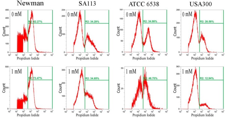
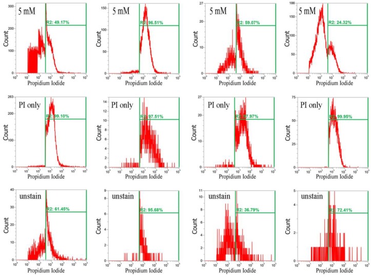
Figure 4.
Analysis of propidium iodide (PI) staining in S. aureus. Four S. aureus strains were grown in MHB supplemented with CuO-NP concentrations of 0–5 mM for 3 h and the cells were then subjected to PI (red) staining. The PI fluorescence intensity is given in arbitrary units (a.u.) on the X-axis, and the Y-axis indicates cell counts as measured by flow cytometry. PBS buffer-only and unstained cells were used as controls. PI staining is fluorescent red color. The data are representative of two separate experiments.


