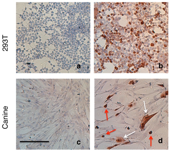
Figure 6.
Sox9 immunostaining of canine and HEK293T cell monolayers. (a) HEK293T and (c) canine MSC in the absence of transfection with MC-CAG.Sox9 vector, no endogenous Sox9 expression is apparent in these culture. (b) HEK293T and (d) canine MSCs MC-CAG.Sox9 vector Sox9 transfected cells showing positive staining for Sox9 protein. Nuclear localization can be observed Sox9 (red arrows), while others show nuclear and cytoplasmic localisation of Sox9 (white arrows). Scale = 200 µm.

