Figure 3.
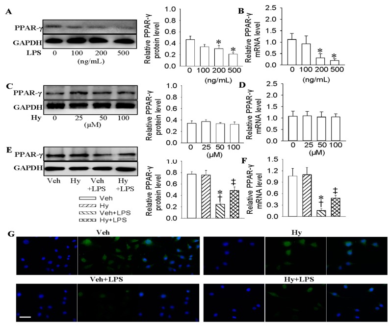
Activation of PPAR-γ mediated the protective role of Hy against LPS-evoked inflammation in HMEC-1 cells. (A) Representative photos showing the effects of various concentrations of LPS (0, 100, 200 and 500 ng/mL for 48 h) on PPAR-γ protein levels in HMEC-1 cells; (B) Effects of various concentrations of LPS (0, 100, 200 and 500 ng/mL for 48 h) on PPAR-γ mRNA levels in HMEC-1 cells; (C) Effects of different doses of Hy (0, 25, 50 and 100 μM for 48 h) on PPAR-γ protein levels in HMEC-1 cells; (D) Effects of different doses of Hy (0, 25, 50 and 100 μM for 48 h) on PPAR-γ mRNA levels in HMEC-1 cells; (E) Representative photographs showing the effects of Hy (100 μM) on PPAR-γ protein expressions in LPS (500 ng/mL)-challenged HMEC-1 cells. The HMEC-1 cells were pretreated with Hy for 6 h before LPS incubation for another 48 h; (F) Effects of Hy (100 μM) on PPAR-γ mRNA expressions in LPS (500 ng/mL)-challenged HMEC-1 cells; (G) Immunofluorescence staining showing the effects of Hy (100 μM) on PPAR-γ expressions in HMEC-1 cells response to LPS (500 ng/mL), scale bar = 50 μm. Values are mean ± S.D. * p < 0.05 vs. 0 ng/mL or Veh, † p < 0.05 vs. Hy, ‡ p < 0.05 vs. Veh + LPS. n = 4 for each group. LPS, lipopolysaccharide; PPAR-γ, peroxisome proliferator-activated receptor γ.

