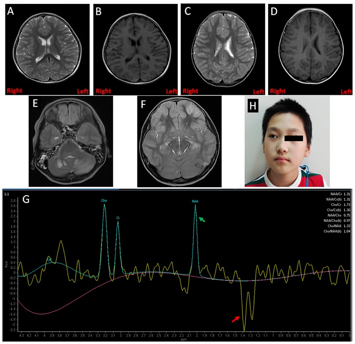
Figure 1.
Clinical features of the patients. (A,B) Magnetic resonance imaging (MRI) showed a mildly enlarged bilateral ventricle in Case 1; (C,D) A mildly enlarged left ventricle was found in Case 2 by MRI; (E,F) Several parts of the brain (cerebellum, frontoparietal, basal ganglia, and thalamus) in Case 4 had symmetrical patchy abnormal signals; (G) Magnetic resonance spectroscopy (MRS) revealed the brain of Case 4 had an abnormal increased lactate peak and reduced N-acetyl aspartate (NAA) peak, which is marked by the red and green arrows, respectively; (H) The facial picture of Case 4 with a strabismic right eye.

