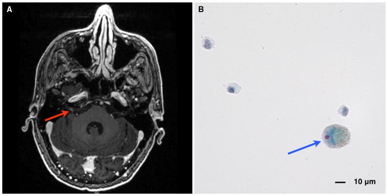
Fig. 2.
a MRI brain, axial T1 with contrast, showing abnormal contrast enhancement of the right internal auditory canal (red arrow), consistent with cranial nerve involvement by leptomeningeal disease from metastatic melanoma. b CSF cytology (Papanicolaou stain) showing enlarged cell (blue arrow) with enlarged, eccentric nucleoli and intracytoplasmic pigment consistent with metastatic melanoma

