Figure 2.
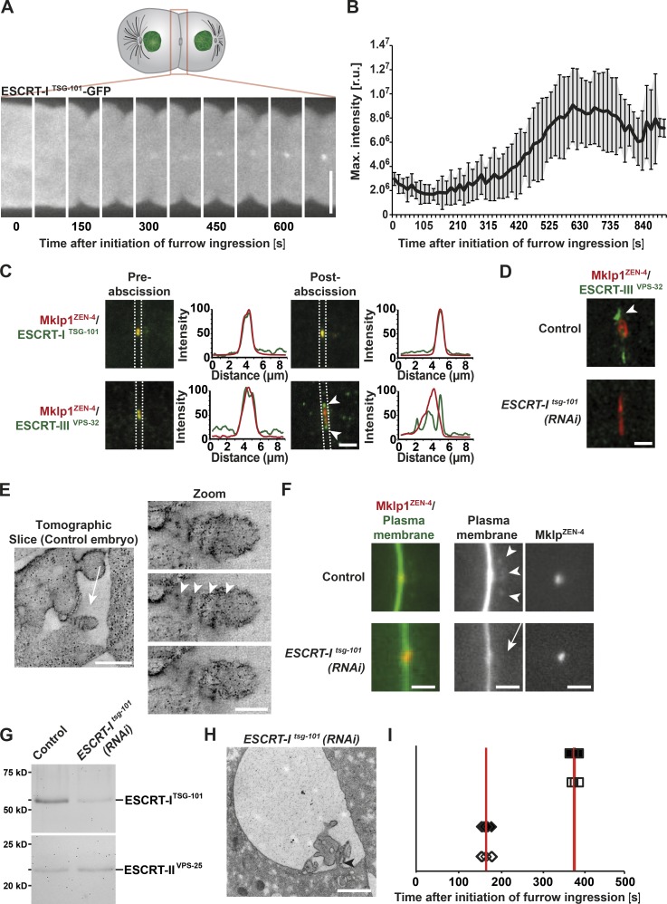
The ESCRT machinery promotes membrane internalization after abscission. (A) Representative images of ESCRT-ITSG-101 accumulation at the intercellular bridge by fluorescence light microscopy. Bar, 10 µm. (B) Quantification of ESCRT-ITSG-101 accumulation. Error bars show SD. r.u., relative units. n = 10. (C) Confocal images showing the accumulation of MKLP1ZEN-4 together with ESCRT-ITSG-101 or ESCRT-IIIVPS-32 before and after abscission. Arrowheads indicate the distinct accumulation of MKLP1ZEN-4 and ESCRT-IIIVPS-32 after abscission. Line scan analysis within the boxed regions was used to measure the distributions of ESCRT components relative to ZEN-4 in each image. n = 6 embryos for each condition. Bar, 2 µm. (D) STED microscopy indicating the accumulation of ESCRT-IIIVPS-32 in control (arrowhead) and tsg-101(RNAi) embryos. n = 6 embryos for each condition. Bar, 1 µm. (E) Tomographic slice of a midbody remnant showing membrane internalization (left, arrow; bar, 500 nm) and a series of tomographic slices of the same membrane bud (right; bar, 100 nm). The spacing of the filaments is indicated (arrowheads). (F) Fluorescence light microscopy of the cleavage furrow of a control (top) and a tsg-101(RNAi) embryo (bottom). Membranes and midbodies were visualized by expressing GFP::PH and mCherry-MKLP1ZEN-4, respectively. Membrane internalization (arrowheads) is visible in the control but absent in the tsg-101(RNAi) embryo (arrow). Bars, 2 µm. n = 12. (G) Immunoblot analysis of control and TSG-101–depleted whole animals using antibodies directed against TSG-101 and VPS-25 (Schuh et al., 2015), a component of the downstream ESCRT-II complex. n = 3. (H) Representative electron micrograph of a midbody remnant in a tsg-101(RNAi) embryo taken 1,100 s after initiation of furrow ingression. The connection of the intercellular bridge to one of the daughter cells is indicated (arrowhead). Bar, 1 µm. n = 3 embryos. (I) Timing of abscission events during the first embryonic division in control and TSG-101–depleted embryos. The start of cleavage furrow ingression is defined as the 0 s time point. Cleavage furrow closure (open diamonds, control; closed diamonds, TSG-101–depleted embryos) and microtubule disassembly (open squares, control; closed squares, TSG-101–depleted embryos) are shown. Red lines highlight the mean time point for each. n = 6 embryos for each condition.

