Figure 3.
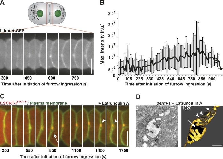
Polymerized actin is required to complete cytokinetic abscission. (A) Representative still images taken of a single embryo show the accumulation of actin at the intercellular bridge by fluorescence light microscopy using a LifeAct-GFP probe. (B) Quantification of actin accumulation after onset of furrow ingression. Error bars show SD. r.u., relative units. n = 15. (C) Representative still images of a latrunculin A–treated embryo during the late stages of cytokinesis. The drug was added after completion of furrow ingression. Localization of the midbody marker ESCRT-ITSG-101 is indicated (arrowheads). A membrane opening (arrow) is visible next to the midbody. n = 12/15. (D) Tomographic slice (left) and 3D model (right) of a high-pressure frozen latrunculin A–treated embryo. The membrane opening next to the intercellular bridge and a fragmentation of the midbody are indicated by an arrow and arrowheads, respectively. The membrane of the intercellular bridge is highlighted in gold. Bars: (A and C) 10 µm; (D) 500 nm.

