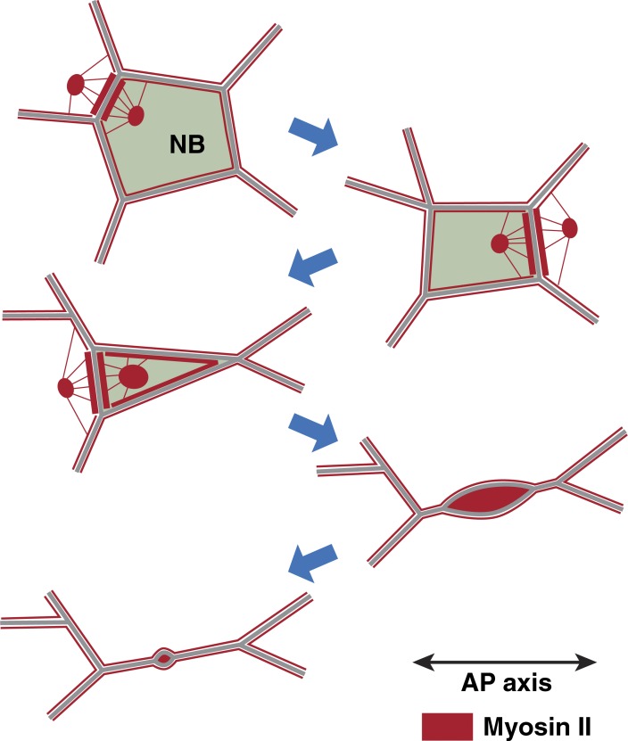
Figure 10.
Model of NB ingression. A typical example of the organization of junctional and medial myosin (red) during five stages of progressive apical constriction in an NB is illustrated. During early ingression (stages 1–3), medial myosin in the NB and neighboring cells flows toward AP NB edges, which become myosin enriched and disassemble. During late ingression (stages 4–5), progressively stronger pulses of medial myosin fuse with DV NB edges, which subsequently disassemble.

