Figure 9.
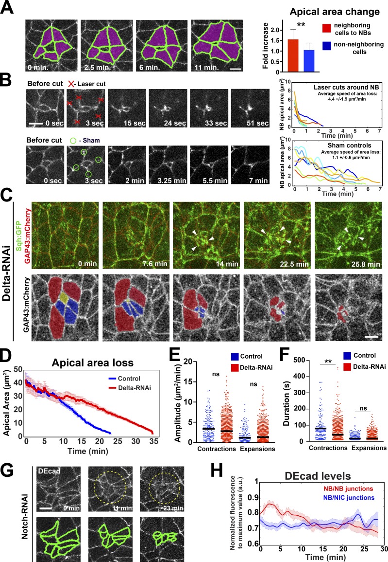
Cell contact and apical expansion of NICs adjacent to NBs are required for normal ingression. (A) NICs (purple) next to NBs expand apically 1.56 ± 0.47–fold during the last 25 min of ingression, in contrast to temporally matched NICs not in contact with NBs (1.05 ± 0.34). n = 25 cells per condition. **, P = 9.8 × 10⁻³ (KS test). (B) Mechanical uncoupling of NBs from neighbors by laser ablation increases ingression speed (top panels and plot) compared with sham-irradiated controls (bottom panels and plot). Each line represents the apical area of one NB after laser/sham cut. Time 0 depicts the apical area before cut. n = 8–9 cells per condition. (C) Cluster of NBs in Delta-RNAi embryos expressing Sqh::GFP (green) and GAP43::mCherry (red). Bottom panels show the same cluster with pseudocolored cells (yellow, blue, and red) ingressing sequentially. Myosin coalesces into foci (arrowheads) during late ingression. (D) Mean apical area loss in control (water-injected Sqh::GFP GAP43::mCherry; 22 NBs, four embryos) and Delta-RNAi embryos (58 NBs, three embryos). (E and F) Amplitude and duration of apical contractions and expansions during ingression in control and Delta-RNAi embryos. Median amplitudes for control/RNAi (E, squared micrometers/minute): contractions, 3.37/2.78; ns (not significant), P = 0.08; expansions, 1.15/1.31; ns, P = 0.509. Median durations for control/RNAi (F, seconds): contractions, 61.57/41.24; **, P = 0.001; expansions, 17.7/17.8; ns, P = 0.6 (KS test). 154–874 events per condition. (G) Cluster of ingressing NBs in Notch-RNAi. Cells are outlined by ubi-DEcad::GFP. DEcad is down-regulated between neighboring NBs (segmented in bottom panels). Bars, 5 µm. (H) DEcad::GFP levels at NB–NB junctions (Notch-RNAi) decrease during ingression, in contrast to NB–NIC junctions in controls. n = 15–16 junctions per condition. Data presented are means ± SEM. a.u., arbitrary units.

