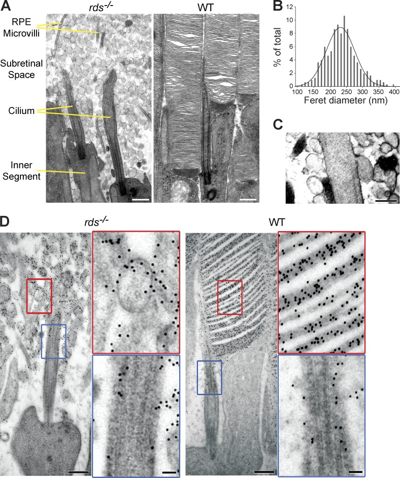
Figure 1.
Ultrastructural analysis of subretinal vesicles in rds−/− mice. (A) EM images of retinal cross sections from rds−/− (left) and WT (right) mice at P21. (B) Gaussian histogram of Feret diameters for extracellular vesicles in rds−/− retinas (n = 250 pooled from two animals). Means ± SD are shown. (C) An example of extracellular vesicle budding from the ciliary membrane of an rds−/− rod. (D) Immunogold labeling of rhodopsin (4D2 mAb) in the distal cilium and extracellular vesicles of a rod photoreceptor in rds−/− and WT retina. Insets show magnified areas marked by red and blue rectangles. Bars: (A and D, main images) 500 nm; (C) 200 nm; (D, insets) 100 nm.

