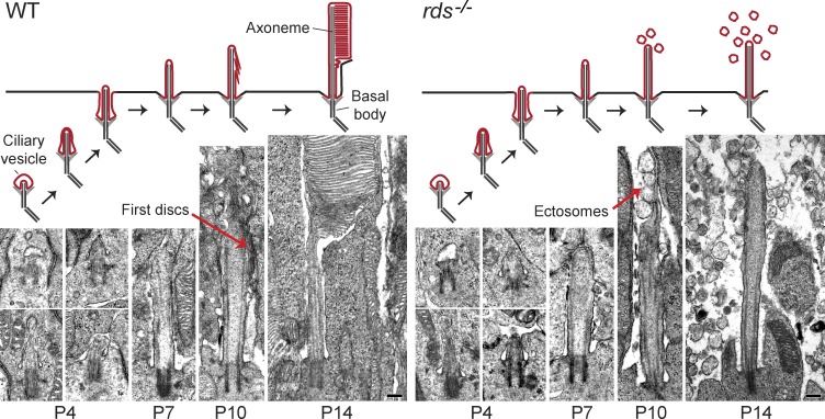
Figure 3.
Developmental stages of the photoreceptor cilium in WT and rds−/− mice. EM images showing the progression of rod outer segment morphogenesis at the indicated postnatal ages for WT and rds−/− retinas. At P10, red arrows highlight the first discs that form at the end of the cilium in WT rods, as opposed to ectosomes that form in rds−/− rods. Schematic representations of each developmental stage are shown above their corresponding images. Bars, 200 nm.

