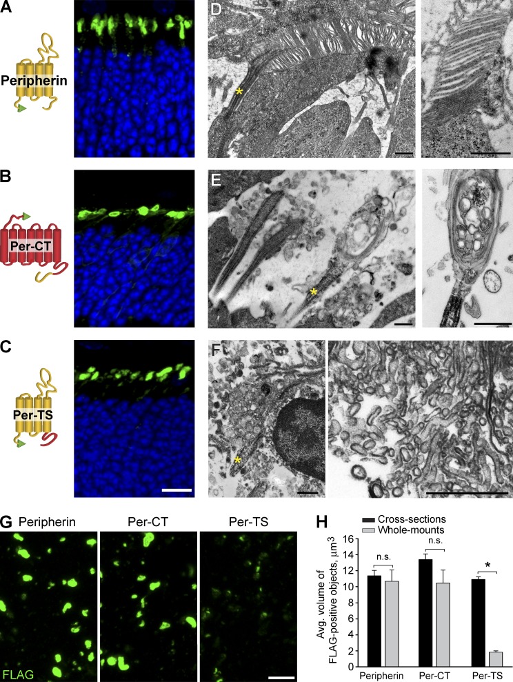
Figure 5.
Transfection of FLAG-tagged recombinant peripherin constructs into rods of rds−/− mice. (A–C) FLAG immunostaining in cross sections of retinas transfected with peripherin (A), Per-CT (B), and Per-TS constructs (C). A cartoon representation of each construct is shown on the left side of each corresponding panel with peripherin sequences shown in yellow, rhodopsin sequences shown in red, and the FLAG tag shown in green. Nuclei are stained with Hoechst (blue). (D–F) EM images of cilia from rods transfected by peripherin (D), Per-CT (E), and Per-TS (F) constructs. Yellow asterisks mark cilia. (G) FLAG immunostaining of retinal wholemounts transfected by each construct after RPE detachment. Bars: (A–C and G) 10 µm; (D–F) 500 nm. (H) Mean volumes of FLAG-positive objects for each construct in retinal cross sections and wholemounts. The data are averaged from at least six individual retinas of each type and are shown as SEM. *, P = 0.009; n.s., not significant. Student’s t test.

