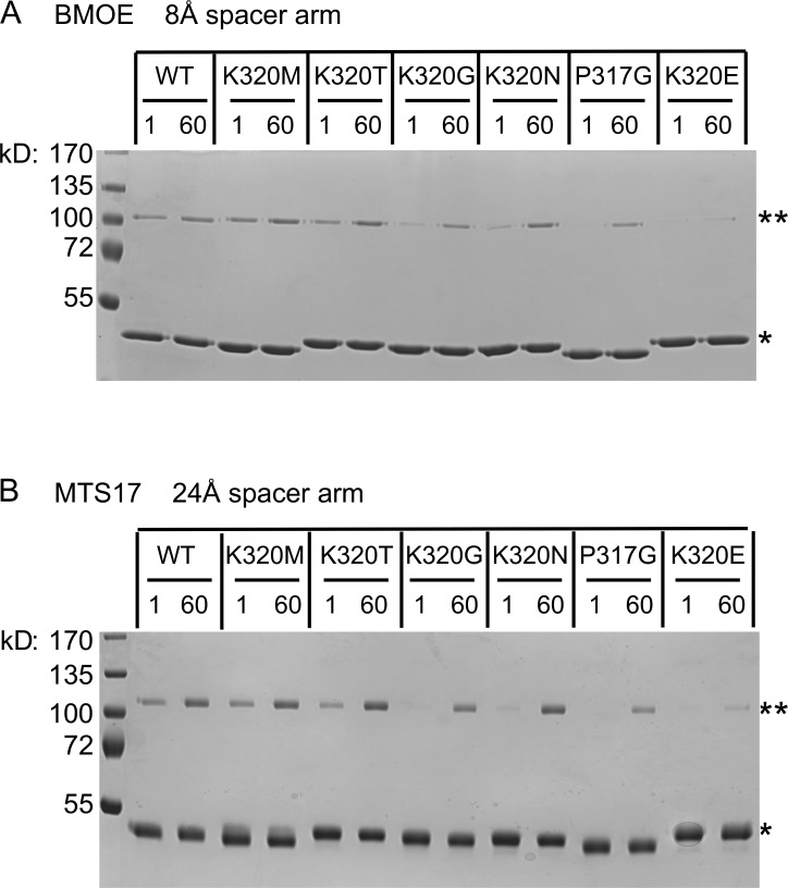
Figure 4.
Cross-linking confirms crossover defects for a subset of mutant variants. Each of the indicated cytoDATL variants was incubated at RT for either 1 min or 60 min in the presence of GMPPNP and then subjected to 20 s of cross-linking with either BMOE (8 Å spacer arm; A) or MTS17 (24 Å spacer arm; B). The structure of each cross-linker is shown to the right. Cross-linked dimers were resolved by SDS-PAGE and visualized with Coomassie blue. The single asterisk marks the monomer, and the double asterisk marks the cross-linked dimer. All variants had the G343C substitution. Concentrations before cross-linker addition were 2 µM CytoDATL and 1 mM nucleotide. The data shown are representative of at least two independent experiments. WT, wild type.

