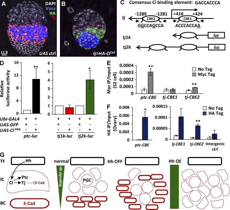
Figure 9.
Hh signaling activates tj transcription via Ci. (A and B) Late-L3 control (ctrl; A) and tj>HA-CiCell gonads (B) with Tj (red, ICs), Vasa (blue, PGCs), DAPI (gray, DNA), and HA (green). Bar, 10 µm. (C) Luciferase expression driven by the 1 kb (tj1k) and 2 kb tj promoters (tj2k) carrying Ci-binding element 1 (CBE1), and both CBE1 and CBE2, respectively. (D) Luciferase reporter assay. S2 cells were transiently transfected with ptc, tj1k, or tj2k luciferase reporter plus ubi-GAL4 together with UAS-GFP or UAS-Ci-PKA. The luciferase activity of ubi-GAL4 & UAS-GFP was set at 1. (E and F) ChIP analysis of Ci binding in S2 cells (E) and in 1-d-old ovaries (F). The chromatin from S2 cells with or without expressing Myc-Ci-PKA and ovaries with or without expressing HA-Ci-PKA was precipitated with antibodies against Myc and HA, respectively. Coprecipitated DNA was analyzed by qPCR using primers against positions containing CBE of the ptc gene, CBE1, and CBE2 of the tj gene. The amplicon for the intergenic region was used as a negative control. Statistical differences in D–F were analyzed by two-tailed t-test. Error bars represent SD in D and SEM in E and F. *, P < 0.05; **, P < 0.001. (G) Hh signaling controls Tj via Ci to suppress E-cadherin (E-Cad) for IC fate. BC, basal cell; OE, overexpression.

