Figure 4.
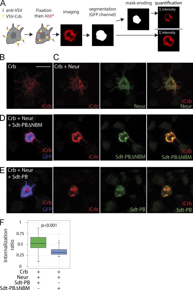
Neur promotes the internalization of Crb via Sdt. (A) Crb internalization assay. S2R+ cells transfected with VSV-Crb were pulsed with anti-VSV antibodies, briefly chased, and then fixed and stained to detect both surface-bound and internalized anti-VSV antibodies (iCrb signal). After image segmentation to detect cell contours (using the GFP signal), an internalization ratio was calculated for each transfected GFP+ cell as the ratio between intracellular and total anti-VSV signals. (B–E) Representative S2R+ cells showing iCrb (red), Flag-tagged Neur (green in C), Flag-tagged Sdt (green in D and E), and GFP (blue). In the absence of Sdt (B and C), iCrb localized mostly into intracellular dots independently of Neur. While iCrb colocalized with Sdt-PB into intracellular dots (E), it colocalized with Sdt-PBΔNBM at the cell cortex. Bar, 10 µm. (F) Box plots showing the internalization ratios measured as described in A. Sdt-PBΔNBM (n = 22 cells), but not Sdt-PB (n = 45), was able to stabilize VSV-Crb at the cell surface in the presence of Neur. A Shapiro test (for normality) and a Wilcoxon rank sum test (for statistical significance) were performed.

