Figure 7.
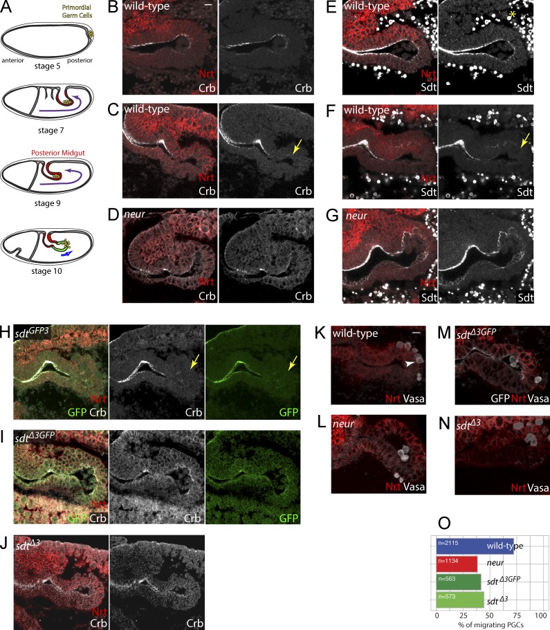
Epithelial remodeling by the Neur-mediated regulation of Crb. (A) PGCs (yellow) form at the posterior pole of the embryo (stage 5) and adhere to the surface of the midgut primordium as it invaginates at stages 7–9. PGCs located in the lumen of the PMG migrate across the epithelium at stage 10. (B–G) Crb (white, B–D; Nrt, red) and Sdt (white, E–G; note that anti-Sdt cross-reacted with yolk granules; yellow asterisk) were detected apically all along the PMG at stage 9 (B and E). At stage 10, Crb (C) and Sdt (F) were no longer detected in distal PMG cells (yellow arrows). In neur mutants, both Crb and Sdt remained detectable at the apical cortex of distal PMG cells (D and G). (H–J) Crb (white) and Sdt-GFP3 (green) were not detected at the apical cortex of distal PMG cells (yellow arrows) in stage 10 sdtGFP3 embryos (H), whereas both Crb and SdtΔ3-GFP (green) remained apical in sdtΔ3GFP embryos (I), indicating that the neur-dependent down-regulation of Crb required the NBM-containing isoforms of Sdt. Similarly, Crb (white) remained apical in distal PMG cells in sdtΔ3 mutant embryos (J). (K–O) PGC migration was quantified at stage 10 by counting the number of PGCs (arrowhead; Vasa, white in K–N) in the lumen, within the epithelium, or inside the embryo (Nrt, red; SdtΔ3-GFP, white; also shown in M). Migrating PGCs correspond to the last two categories (n = 563–2,115 PGCs; for each genotype, n is the total number of PGCs scored from >30 embryos). PGC migration across the PMG epithelium appeared to be similarly delayed in neur, sdtGFP3, and sdtΔ3 mutant embryos (O). Bars, 10 µm.

