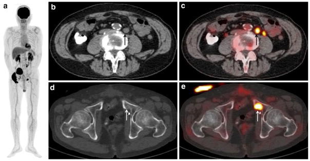
Fig. 2.
A 66-year-old man with bladder cancer (cT3N1) after neoad-juvant chemotherapy and radical cystectomy, who developed lymph node and bony metastases. a MIP of FDG-PET shows two areas of abnormal FDG uptake in the pelvis. b CT of FDG-PET/CT and c PET/CT show moderate FDG uptake (SUVmax: 7.2) corresponding to a 1.5-cm swollen para-aortic lymph node (arrow), suggesting lymph nodal recurrence. A tiny FDG uptake which is located to the left of that nodal recurrent lesion is physiologic excretion in the left ureter. d CT of FDG-PET/CT and e PET/CT show intense FDG uptake (SUVmax: 13.0) corresponding to the mild sclerotic change of the left pubic bone (arrow), suggesting bone metastasis. The detection of this bony metastasis by only (d) CT is difficult

