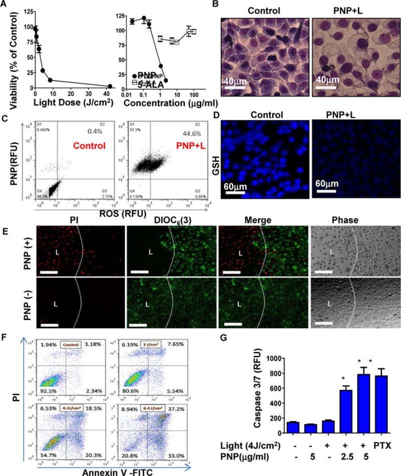
Figure 3. In vitro antitumor efficacy and cytotoxic mechanisms of PNPs against bladder cancer cells.

(A) left: the viability of 5637 bladder cancer cells at 24 hours post PNP treatment and illumination with different doses of light (Pyropheophorbide a: 2 μg/ml for 2 hours) and different PNP and 5-ALA concentrations (right) (Light dose: 4.2 J/cm2). (B) Cell morphology changes at 3 hours post PNP-mediated photodynamic therapy. (Hema3™, 1000× oil). (C) Intracellular ROS production and (D) Glutathione (GSH) levels in 5637 cells upon photodynamic therapy. (E) Mitochondria membrane potential (DiOC6(3): green) and cell integrity/viability (PI : red) 24 hours post treatment. Cells were incubated with DiOC6(3) and PI for 20 minute. DiOC6(3) low referred to loss of membrane potential, while PI + (red) stained dead cell nucleus. Bar=150 μm. (F) Apoptosis/necrosis assay, and (G) caspase 3/7 activation of 5637 cells at 24 hours post photodynamic therapy. PTX (1μg/ml) treated groups were served as positive control. (PI+/Annexvin V+ : late apoptosis; PI-/Annexvin V+: early apoptosis; PI+/Annexvin V− : Necrosis). (n=3, t-test, * p<0.05).
