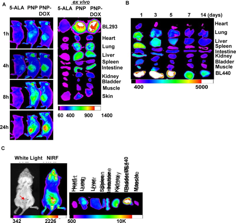
Figure 6. NIRF imaging of PDX mice models bearing subcutaneous and orthotopic bladder cancer.

(A) In vivo NIFR imaging of NSG mice bearing subcutaneous PDX BL293 up to 24 hours after intravenously administration of 5-ALA (100 mg/kg), PNPs (Pyropheophorbide a 5 mg/kg), and PNP-DOX (Pyropheophorbide a 5mg/kg and DOX 2.5 mg/kg). Right: ex vivo NIFR imaging for BL293 tumors and other major organs. (5-ALA induced protoporphyrin IX ex/em = 633/650–710nm; PNP ex/em = 680/690nm. Kodak imaging system 650/700nm) (B) Biodistribution and tumor retention of PNP-DOX at different time points after injection. (C) The NIRF imaging of mice bearing orthotopic BL440 PDX model (red arrow) 24 hours after the administration of PNP. (Left panel: In vivo whole mouse imaging; Right panel: ex vivo imaging)
