Figure 6.

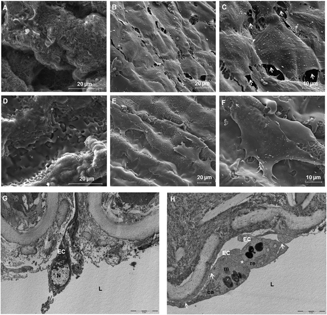
Porcine recellularized arteries after 5 days in culture. Representative scanning electron microscopy (SEM) images of the luminal side of the arteries: (A) decellularized artery with no cells, (B and C) non polymer-coated recellularized arteries, white arrows are pointing out gaps between cells, (D) polymer-coated decellularized arteries, (E and F) polymer-coated recellularized arteries showing the formation of a continuous cell monolayer. (Magnification; (A,C,D and F) x4000, (B and E) x1600). (G and H) Transmission electron microscopy (TEM) analyses of sections cut in perpendicular to the long axis of the artery of (G) native arteries and (H) polymer-coated recellularized arteries. (H) White asterisk show cell adhesion between two cells and white arrows point out adhesion between the cell and the luminal side of the vessel. L: lumen, EC: endothelial cells, N: nucleus, ca: caveolae, er: rough endoplasmic reticulum, m: mitochondria, scale bar indicates: (A) 10 μm, (B) 5 μm.
