
Figure 1. α‐Synuclein overexpression alters spine density and dynamics in vivo .
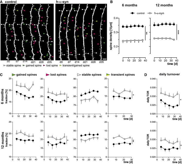
- Representative in vivo two‐photon recordings of eGFP‐labeled apical tuft dendrites in the somatosensory cortex in h‐α‐syn and control animals. Arrowheads mark representative spines that were stable (white, present > 7 days), newly formed (green), or lost (magenta). Gained spines that do not stabilize (yellow/green, present < 7 days) are defined as transient. Scale bar, 5 μm.
- Spine density is reduced in both 6‐ (**P = 0.0012) and 12‐month‐old (***P = 0.0001) h‐α‐syn animals.
- The fractions of both gained (P 6 months = 0.0527; P 12 months = 0.0678) and lost spines (*P 6 months = 0.0461; *P 12 months = 0.0206) are elevated in h‐α‐syn mice compared to controls; the fraction of transient spines is significantly higher (*P 6 months = 0.0315; **P 12 months = 0.0017).
- Consequently, the daily turnover ratio (TOR) is significantly increased in both 6‐ and 12‐month‐old h‐α‐syn mice (*P 6 months = 0.0491; *P 12 months = 0.0338).