
Figure 2. NELF‐E is rapidly recruited to DNA damage sites to induce transcriptional silencing.
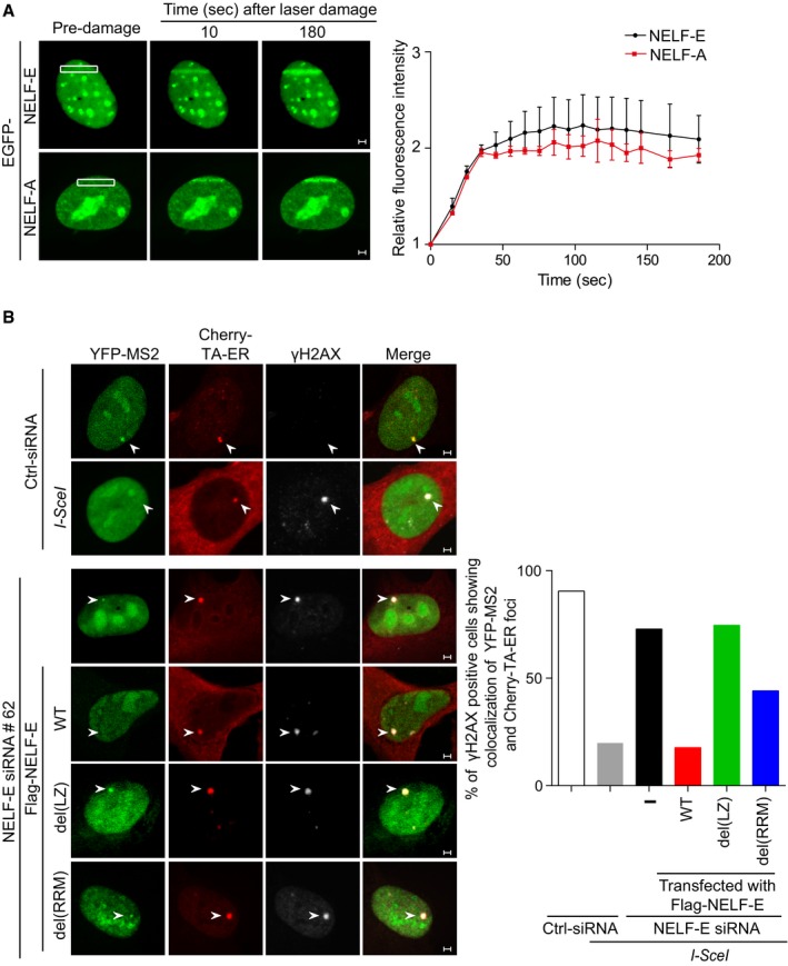
- Representative time‐lapse images showing the localization of EGFP‐NELF‐E and EGFP‐NELF‐A after laser microirradiation targeted to a particular region marked by white rectangle. Scale bar, 2 μm. Graph on the right shows fold increase in the relative fluorescence intensity at laser‐microirradiated sites. Results shown are typical of four independent experiments and represent 40 different cells. Error bars indicate standard error of the mean (SEM).
- NELF‐E recruitment to DSB is critical for transcriptional silencing. NELF‐E‐deficient U2OS‐TRE‐I‐Sce‐19 cells were transfected with pCherry‐tTA‐ER, pYFP‐MS2, and pCMV‐NLS‐I‐SceI plasmids along with either of the indicated constructs expressing Flag fused to NELF‐EWT, NELF‐Edel(LZ), or NELF‐Edel(RRM). Cells were immunostained with γH2AX (gray). Graph displays the percentage of γH2AX‐positive cells that show colocalization of YFP and Cherry foci. White arrowheads mark the location of the MS2 reporter cassette. Data represent the mean of two biological repeats. Scale bar, 2 μm.