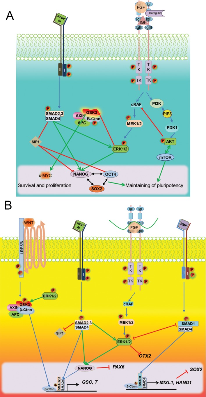
Fig.1.

Molecular interplay of FGF and NODAL signaling in maintenance of pluripotecy in hPSCs and mesendodermal differentiation. A. Self-renewal of hESCs depend on activation of both fibroblast growth factor (FGF) and activin/nodal signals. Activin/Nodal signals bindsto typeI/II receptors. Hetrodimerization of receptors and their phosphorylation results in activation of R-Smads (SMAD2/3) and their binding to the co-SMAD (SMAD4). Internalization of the SMAD protein complex to the nucleus activates expression from NANOG directly. Indirectly, this could sustain the core pluripotency network and expressions of OCT4 and SOX2. SMAD proteins also inhibit expression of the SMAD interacting protein (SIP) which has a negative effect on OCT4 and NANOG expression, and could enhace neuroectodermal differentiation. FGF activation, by binding of their ligands [FGF, insulin-like growth factor (IGF) and heregulin] to tyrosine kinase receptors, results in phosphorylation and activation of phosphatidylinositol 3-kinase (PI3K) and cRAF. Activation of PI3K in turn activates AKT, which is involved in mediating inhibition of apoptosis and stimulation of cell proliferation, specifically via mTOR signaling in human pluripotent stem cells (hPSCs). Activation of cRAF activates mitogen-activated protein kinase MEK/ERK signaling that controls cellular processes such as survival and differentiation, and can maintain NANOG expression. A moderate signal of internal glycogen synthase kinase-3 (Gsk3) in hPSCs is needed for cell proliferation via c-Myc expression and B. Combined signals of mesendoderm specification. Activation of p-SMAD2/3 downstream of activin/nodal external signals reinforces the expression of NANOG and primitive streak specific genes (GSC and T). The high expression level of the ERK signaling, downstream of FGF, helps to stabilize th expression of β-ctnn and the mesendodermal genes. Phosphorylation of SMAD1 and its dimerization with SMAD4 occurs as a result of BMP binding to its receptors which enhance the expression of the posterior mesoderm and extra-embryonic mesodermal genes (Hand1 and Mixl1). Expressions of neural specific genes (SIP1, PAX6, OTX2 and SOX2) that are inhibited by mesodermal signals are depicted by red lines.
