-
A
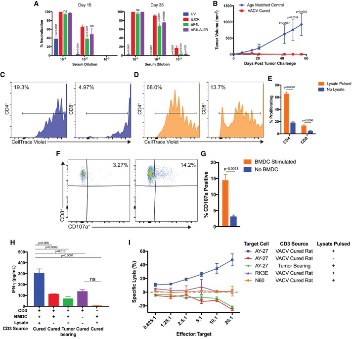
VACV‐neutralizing antibodies were measured in virus‐treated rats 15 and 35 days after implantation (n = 4–5 rats per group).
-
B
Protection from subcutaneous tumor challenge after virus‐induced tumor clearance. AY‐27 cells were implanted in the flanks of cured rats (n = 6) and naïve age‐matched control rats (n = 4).
-
C–E
T‐cell proliferation after co‐culturing with bone marrow‐derived dendritic cells (BMDCs). CD4+ and CD8+ cells were co‐cultured with BMDCs and proliferation assayed with CellTrace Violet. The representative plots show CD4+ and CD8+ T‐cell proliferation after co‐culture with either mock‐pulsed (C) or with tumor‐lysate‐pulsed BMDCs (D). Panel (E) shows the percentage of CD4+ and CD8+ T cells that proliferated in response to BMDC stimulation (n = 3).
-
F, G
Ex vivo upregulation of CD107a by CD8+ T cells from challenged rats. (F) CD3+ cells were incubated +/− BMDCs for 1 h in the presence of anti‐CD107a antibody, incubated for 5 h with monensin and brefeldin A, and then stained with anti‐CD4 and anti‐CD8 antibodies. Events were gated for viable CD8+ T cells. Panel (G) shows the percentage of CD107a+ CD8+ T cells +/− BMDC stimulation (n = 3).
-
H
IFN‐γ released after 24‐h co‐culture of CD3+ cells with BMDCs (n = 3–5).
-
I
T cells activated ex vivo by tumor‐lysate‐pulsed DCs are cytotoxic. After 6 days of co‐culture with BMDC, CD3+ cells were incubated for 18 h with 10,000 target cells and at different effector‐to‐target ratios. Lysis was determined by LDH assay. RK3E are normal rat kidney cells (n = 2–3 performed in duplicate).
Data information: Mean ± SEM is shown. Two‐way ANOVA followed by Tukey's multiple comparison test was used in (A), (B), and (H). For (A), significance was determined against the ∆
J2R group. Two‐tailed Student's
t‐test was used in (E) and (G).