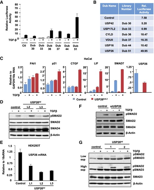
Figure EV1. USP26 enhances SMAD2 phosphorylation and TGF‐β‐mediated transcription.

- Graph representing relative luciferase values obtained from DUB screen. 293T cells were transfected with CAGA‐Luc and indicated DUB pools. Forty‐eight hours later, cells were treated with TGF‐β for 16 h and a luciferase assay was performed. Data are mean ± SD of triplicate samples.
- Table indicates relative luciferase value of each gene tested in (A).
- HaCat cells stably transduced with a hairpin targeting USP26 or vector control were stimulated with TGF‐β for 3 h. PAI1, CDKN1A (p21), CTGF, LIF, SMAD7, and USP26 mRNA levels relative to GAPDH or 18S are shown as evaluated by quantitative real‐time PCR. Data are mean ± SD of triplicate samples.
- 293T cells were stably infected with two independent hairpins (L2 and L3) targeting USP26 and treated with TGF‐β overnight. Whole‐cell extracts were probed with the indicated antibodies.
- 293T cells were stably infected with three independent hairpins (L1, L2, and L3) targeting USP26. USP26 mRNA levels relative to 18S are shown as evaluated by quantitative real‐time PCR. Data are mean ± SD of triplicate samples.
- 293T cells transfected with siRNA targeting USP26 or control vector were treated with TGF‐β overnight. Whole‐cell extracts were probed with the indicated antibodies.
- 293T cells expressing knockdown vectors targeting USP26 or control vector were treated with TGF‐β overnight. Whole‐cell extracts were probed with the indicated antibodies.
Source data are available online for this figure.
