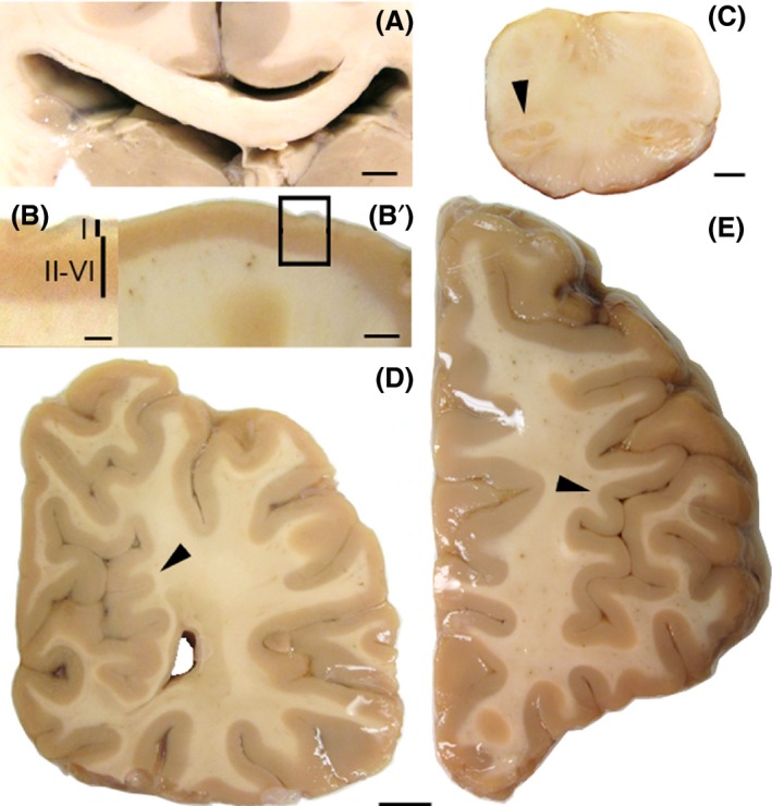
Figure 2.

Thick coronal sections of the brain. (A) Thin corpus callosum and expanded left lateral ventricle. (B and B’) Thin cortex and thin II‐VI layers when compared to the thickened layer I. (C) Small left inferior olive. (D) Gyri crowding (arrowhead) in the medial region of the occipital lobe. (F) Abnormal gyrification (arrowhead) in the lateral frontal lobe. Calibration bars: A: 6 mm, B: 4 mm, C. 2 mm, D–E: 1 cm.
