
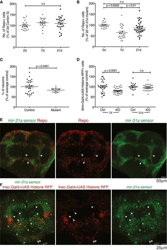
Figure EV1. miR‐31a is expressed in adult progenitor cells that give rise to glia (related to Fig 1).
-
A, BNumber of glia at 2, 7 and 21 days post‐eclosion represented as a percentage of the number in 2‐day‐old flies. Error bars represent SEM. Data were analysed using one‐way ANOVA. (A) Canton S controls. (B) miR‐31a mutants.
-
CSmall significant difference in number of neurons in the central brain in 7‐day‐old adults was observed. Data are represented as a percentage of the average number of neurons in Canton S control animals. Data were quantified with Imaris (Bitplane). Unpaired Student's t‐test was used for analysis. Error bars represent SEM.
-
DAstrocyte numbers (Alrm‐Gal4 > UAS‐Histone‐RFP) in 7‐days and 21‐days controls (Ctrl) and miR‐31a mutants (KO) represented as a percentage of the number in the CS controls. Unpaired Student's t‐test was used for analysis. Error bars represent SEM.
-
EmiR‐31a sensor in a 2‐days post‐eclosion adult brain. miR‐31a activity is indicated by the absence of GFP expression. White arrowheads point to example cells where GFP co‐localizes with anti‐repo (red), indicating low miRNA activity in the mature glia.
-
FmiR‐31a sensor (GFP) expression is excluded from some Insc‐Gal4 > UAS‐Histone‐RFP‐expressing cells (white arrowheads) in the brains of 2‐days post‐eclosion adults.