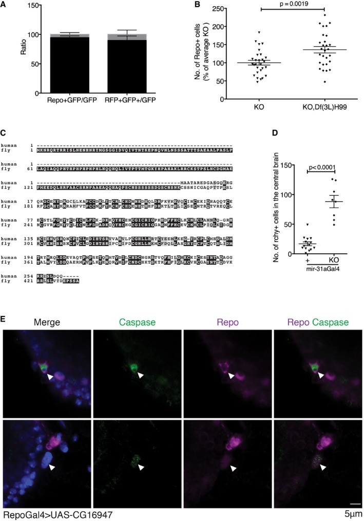
Figure EV2. Rchy1, the target of miR‐31a, causes death of glia by apoptosis (related to Figs 2 and 3).

- Left panel: number of repo‐expressing and lineage‐tagged GFP+ cells. Right panel: lineage‐tagged GFP+ cells with active repo‐Gal4. Data are presented as a ratio of the total number of GFP cells in the central brain. Error bars represent SEM.
- Number of glia in miR‐31a mutants at 7 days with and without Df(3L)H99. Introducing Df(3L)H99 prevented glial loss. Data were analysed with an unpaired Student's t‐test. Error bars represent SEM.
- Alignment of fly and human Rchy1 proteins showing regions of sequence similarity. The antibody to human Rchy1 was raised against a peptide containing residues 87–167.
- The number of Rchy1‐expressing cells in the central brain was greater in the miR‐31a mutants than in Canton S controls (88 ± 0.4 vs. 17 ± 3). Data were analysed with an unpaired Student's t‐test. Error bars represent SEM.
- Activated caspase‐3‐positive (green) glial cells stained with anti‐repo (purple) are observed in the brains of 2‐days post‐eclosion adult repo‐Gal4 > UAS‐CG16947 brains. White arrowheads point to anti‐repo‐positive cells that are activated caspase‐3‐positive.
