
Figure 1.
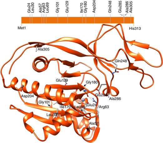
Schematic and structural representation of the ASPA monomer with the location of the amino acid residues addressed in this study. The structural image was generated using the crystal structure of ASPA (PDB ID: 2O4H), obtained using the UCSF Chimera package. Chimera is developed by the Resource for Biocomputing, Visualization, and Informatics at the University of California, San Francisco (supported by NIGMS P41‐GM103311).