
Figure 1.
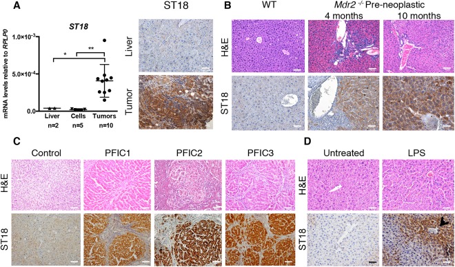
ST18 is induced by inflammatory cues in hepatoblasts. (A) Left: Quantitative reverse‐transcription PCR analysis of ST18 mRNA levels in adult liver in cell lines derived from mouse liver progenitors (including E14.5 shp53‐Myc, E14.5 shp53‐RasV12, E14.5 Myc‐RasV12, E18.5 shp53‐Myc‐RasV12, and E18.5 shp53‐RasV12) and in subcutaneous tumors derived from the same cells. *P = 0.002. **P = 0.0462. Right: ST18 is expressed in subcutaneous hepatoblast‐derived tumors but not normal liver. (B) Hematoxylin and eosin (H&E) staining shows portal inflammatory infiltrates in preneoplastic Mdr2 −/− livers at the indicated ages. Staining of IHC sections with ST18 antibodies (ST18) shows positivity in Mdr2 −/− but not wild‐type livers. (C) Hepatic lesions and ST18 positivity in liver biopsies from PFIC1, PFIC2, and PFIC3 patients. (D) Mouse liver sections 24 hours after LPS treatment showing irregular hepatocyte arrangement, inflammatory infiltrates, and induction of ST18, in particular nearby blood vessels (arrow) in both the periportal and centrilobular zones.