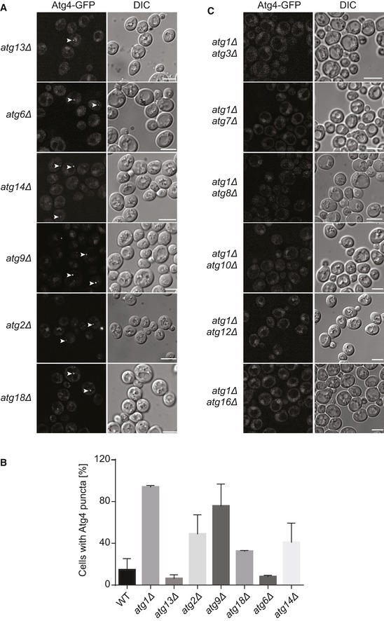
Figure EV2. Atg4 association with the PAS does not require components of the Atg1 complex, Atg9 cycling system and PI3K complex.

- Fluorescence microscopy images showing the subcellular localization of Atg4‐GFP in atg13∆ (SAY030), atg2∆ (SAY109), atg9∆ (SAY016), atg18∆ (SAY017), atg6∆ (SAY013), and atg14∆ (SAY110) strains analyzed as in Fig 1. White arrows highlight Atg4‐GFP puncta. DIC, differential interference contrast. Scale bars, 5 μm.
- Percentage of cells, in the experiments shown in panel (A), that display an Atg4‐GFP punctate structure. Data represent the average of three independent experiments ± SD.
- Subcellular localization of Atg4‐GFP in double knockout cells lacking Atg1 and components of the conjugation systems leading to the formation of Atg8‐PE: atg7∆ (SAY032), atg3∆ (SAY031), atg8∆ (SAY033), atg10∆ (SAY035), atg12∆ (SAY134), and atg16∆ (SAY135). Cells were grown and imaged as in Fig 1. DIC, differential interference contrast. Scale bars, 5 μm.
