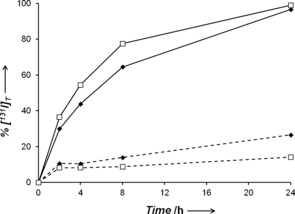
Figure 2.

Release of free 131I into the supernatant of cell culture medium at various times after incubation with 1‐[131I] (♦) or 2‐[131I] (□), in the absence (dashed lines) or presence (solid lines) of MCF‐7 breast cancer cells. Percentages were determined by HPLC peak integrals.
