
Figure 1. STAT3 is phosphorylated on residue Tyr705 during IFNγ/TNFα‐induced muscle wasting.
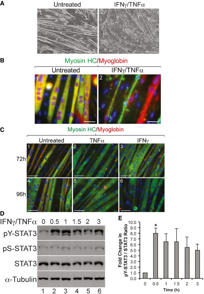
C2C12 cells were grown to confluency then induced for differentiation for 4 days to form fully differentiated myotubes. These myotubes were exposed or not to IFNγ/TNFα for 72 or 96 h.
- Phase contrast images of cultured C2C12 myotubes treated with or without IFNγ/TNFα for 72 h. Scale bar = 200 μm. Images shown are representative of n = 4 independent experiments.
- Immunofluorescence (IF) images of C2C12 myotubes either untreated or treated with IFNγ/TNFα for 72 h were stained with anti‐myosin heavy‐chain (MyHC; green) and anti‐myoglobin (red) antibodies. Scale bar = 50 μm. Images shown are representative of n = 4 independent experiments.
- IF images of C2C12 myotubes treated for 72 h (panels 1–3) or 96 h (panels 4–6) with either TNFα (panels 2 and 5) or IFNγ (panels 3 and 6). Untreated (panels 1 and 4) and treated myotubes were stained with myosin heavy chain (green) and myoglobin (red) as marker for differentiated muscle cells. Images shown are representatives of n = 2 independent experiments. Scale bar = 50 μm.
- Total extracts of C2C12 myotubes treated with or without IFNγ/TNFα were used for Western blot analysis with antibodies against pY‐STAT3, pS‐STAT3, total STAT3, and α‐tubulin. The blot shown is a representative of n = 4 independent experiments.
- Densitometric quantification of pY‐STAT3 signal relative to total STAT3 signal in panel (D). Data represented as mean ± SEM (n = 4) with *P‐value = 0.0135 by one‐way ANOVA with Dunnett's post hoc test.