
Figure 7. Active NF‐κB pathway is required for the rapid translocation of pY‐STAT3 to the nucleus during IFNγ/TNFα‐induced muscle wasting.
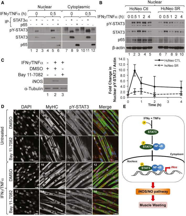
- Nuclear and cytoplasmic fractions prepared from C2C12 myotubes treated with IFNγ/TNFα followed by immunoprecipitation (IP) with either total STAT3, p65, or an IgG control antibody. Western blot analysis was performed using antibodies against total STAT3, pY‐STAT3, and p65. The blot shown is a representative of n = 3.
- (Upper panel) Nuclear and cytoplasmic fractions were prepared from hcNeo control (Ctl) and hcNeo Super Repressor (SR) cells treated with IFNγ/TNFα and used for Western blot analysis with antibodies against pY‐STAT3, total STAT3, p65, and β‐actin. The blot shown is a representatives of n = 2. (Lower panel) Densitometric quantification of nuclear pY‐STAT3 relative to β‐actin. Data are represented as mean ± SD (n = 2).
- Total cell extract from C2C12 myotubes pre‐treated with Bay 11‐7082 or DMSO as a control then treated with IFNγ/TNFα for 24 h were used for Western blot analysis with antibodies against iNOS and α‐tubulin. The blot shown is a representative of n = 2.
- Confocal microscopy images of C2C12 myotubes pre‐treated with Bay 11‐7082 or DMSO as a control, then treated with or without IFNγ/TNFα and stained for pY‐STAT3 (red), MyHC (green), and counterstained with DAPI (blue). Scale bar = 20 μm. Images are representative of n = 1 experiment.
- Model depicting the how STAT3 promotes cytokine‐induced muscle wasting. The cytokines IFNγ and TNFα act synergistically by activating STAT3 via phosphorylation on its Y705 residue. Following the degradation of IκBα (not shown here), pY‐STAT3 translocates to the nucleus as a complex with NF‐κB and upregulates the expression of iNOS, leading to the activation of the iNOS/NO pathway, which in turn promotes muscle wasting.
Source data are available online for this figure.