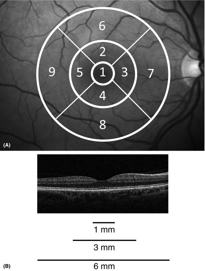
Figure 1.

Optical coherence tomography (OCT) scan and the nine sectors of the ETDRS grid. (A) The nine sectors of the ETDRS grid (right eye). Fovea (sector 1), pericentral ring (sectors 2–5) and peripheral ring (sectors 6–9). (B) Representative horizontal spectral domain Cirrus HD‐OCT (Carl Zeiss Meditec) scan through the fovea from the right eye of a healthy subject.
