Figure 1.

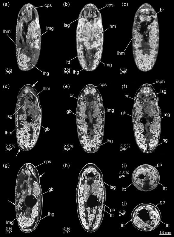
Calliphora vicina, micro‐CT‐based virtual sections of puparia at different times after pupariation (AP), reared at 24°C. The corresponding percentage of time of the total intra‐puparial period (IPP) is given in brackets after each time. (a) 0 hr AP (0% IPP), medial sagittal section. (b) 0 hr AP (0% IPP), dorsal horizontal section. (c) 0 hr AP (0% IPP), medial horizontal section. (d) 6 hr AP (2.5% IPP), lateral sagittal section. (e) 6 hr AP (2.5% IPP), medial horizontal section. (f) 6 hr AP (2.5% IPP), ventral horizontal section. (g) 12 hr AP (5% IPP), medial sagittal section. (h) 12 hr AP (5% IPP), medial horizontal section. (i) 6 hr AP (2.5% IPP), medial cross section of the abdomen. (j) 12 hr AP (5% IPP), medial cross section of the abdomen. br, brain; cps, cephalopharyngeal skeleton; gb, gas bubble; lfg, larval foregut; lhg, larval hindgut; lhm, larval hypodermal muscles; lmg, larval midgut; ltt, larval tracheal trunks; lsg, larval salivary glands; rsph, respiratory horns. Arrows indicate sites where larval‐pupal apolysis has occurred
