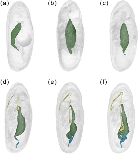
Figure 4.

Calliphora vicina, false‐color 3D‐surface models of puparia at different times after pupariation (AP), reared at 24°C, showing the changes in the adult alimentary canal. (a) 24 hr AP. Note the presence of the gas bubble in the central part of the abdomen. (b) 30 hr AP. (c) 48 hr AP. (d) 72 hr AP. (e) 96 hr AP. (f) 120 hr AP. Foregut shown in yellow, midgut in green and hindgut in blue
