Figure 3.

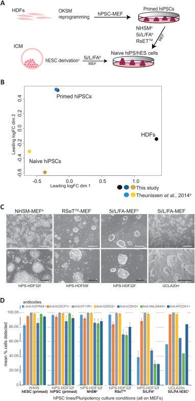
Cell surface antigens are detected on naive state human pluripotent cells. Transcriptional and protein analyses showing the expression of cell surface epitopes on human iPS cells (hiPSCs) and human ES cells (hESCs) cultured in conditions supporting a naive state of pluripotency. (A): Schematic depicting the generation of naive human cell cultures both from lineage primed hiPSCs and blastocyst epiblast cells. Human dermal fibroblasts (HDFs) were reprogrammed to primed hiPSCs in standard human pluripotent stem cell (hPSC)‐MEF culture then coaxed to a naive pluripotent state in NHSMa, RSET, and 5i/L/FAb defined media‐MEF supported culture conditions. Naive state hESCs were derivedc and maintained following the direct culture of preimplantation blastocyst in 5i/L/FA‐MEF conditions. (B): PCA of RNA sequencing data for the primed and naive (5i/L/FA) state hiPSCs from this study and the microarray data extracted from the published report of Theunissen et al. (2014), 38 confirms differential clustering of two parental HDFs (black dot), lineage primed hiPSCs from this study (dark blue dot) and Theunissen et al. (2014), (light blue dot), and naive hiPSCs from this study (dark yellow dot) clustering with the naive hiPSCs from Theunissen et al. (2014) (gold dot). (C): Representative images for naive hiPSC colonies originating from two parental HDF cell lines (HDF32f, HDF55f) following culture in NHSMa‐MEF, RSeT‐MEF, and 5i/L/FAb‐MEF conditions and preimplantation blastocyst‐derived hES naive cell colonies (UCLA20nc) generated directly in 5i/L/FA‐MEF culture conditions. HiPS and hESC naive cell cultures show a domed colony morphology by bright field phase contrast microscopy (BF). (D): Flow cytometric replicate analyses shown graphically for the live cell detection of naive state hiPS‐HDF32f cells maintained on MEFs in NHSMa, 5i/L/FAb, and RSeT culture media, and naive UCLA20n hESCs in 5i/L/FA‐MEF culture, by the monoclonal antibodies anti‐hGPR64 (light blue bars), anti‐hCDCP1 (orange bars), anti‐hF11R (gray bars), anti‐hDSG2 (yellow bars), anti‐hCDH3 (mid blue bars), anti‐hNLGN4X (green bars), anti‐hPCDH1 (dark blue bars), compared with lineage primed WA09 cells (hESC primed) and parental hiPS‐HDF32f cells (hiPSC primed) cultured on MEFs in hPSC medium, against isotype controls (not shown), (n = 3, except UCLA20n in 5i/L/FA n = 2). Error bars depict SEM. Scale bars = 500 µm (white) and 200 µm (black) for BF images. aGafni O et al. Nature 2013;504:282‐286. bTheunissen TW et al. Cell Stem Cell 2014;15:471‐487. cPastor WA et al. Cell Stem Cell 2016;18:323‐329. Abbreviations: hESCs, human ES cells; hiPSCs, human iPS cells.
