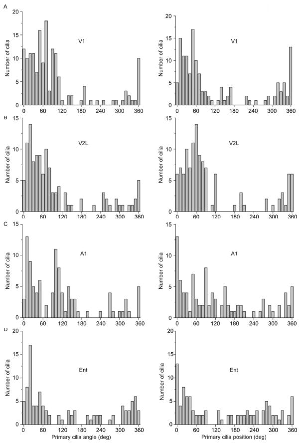
Figure 2.
Neuronal primary cilia alignment in the cortex. (A) Shown are the distribution of angles (°) of primary cilia with respect to the cortical surface (left) and with respect to the soma (right) in area V1. (B) Shown are the distribution of angles (°) of primary cilia with respect to the cortical surface (left) and with respect to the soma (right) in area V2L. (C) Shown are the distribution of angles (°) of primary cilia with respect to the cortical surface (left) and with respect to the soma (right) in area A1. (D) Shown are the distribution of angles (°) of primary cilia with respect to the cortical surface (left) and with respect to the soma (right) in area Ent. Angle: N = 159 cells (V1), 116 cells (V2L), 102 cells (A1), 101 cells (Ent); Position: N = 141 cells (V1), 118 cells (V2L), 102 cells (A1), 101 cells (Ent) from 4 animals.

