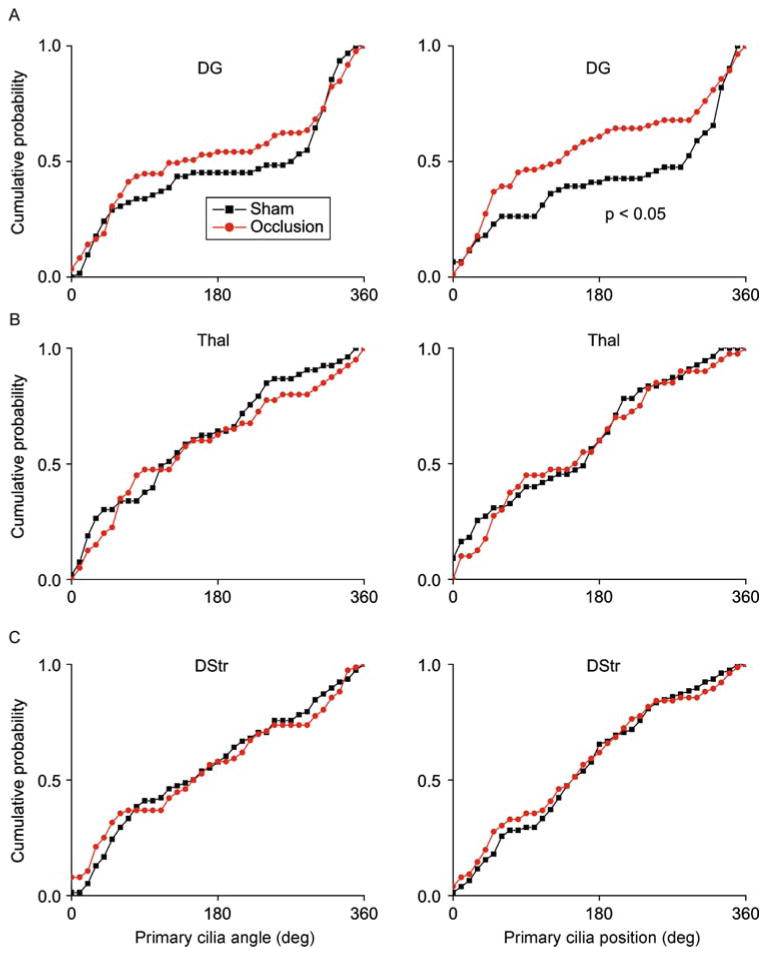
Figure 8.
Internal carotid artery occlusion affected primary cilia alignment in the dentate gyrus but not in striatum or thalamus. (A) Shown are distribution plots of primary cilia angle and position (°) in DG. (B) Shown are distribution plots of primary cilia angle and position (°) in Thal. (C) Shown are distribution plots of primary cilia angle and position (°) in DStr. Two-sample Kolmogorov–Smirnov tests: DG angle: P >0.05, DG position: P = 0.011, N = 61 cells (sham), 85 cells (occlusion); Thal angle: P >0.05, Thal position: P >0.05, N = 55 cells (sham), 40 cells (occlusion); DStr orientation: P >0.05, DStr orientation: P >0.05, N = 78 cells (sham), 76 cells (occlusion).

