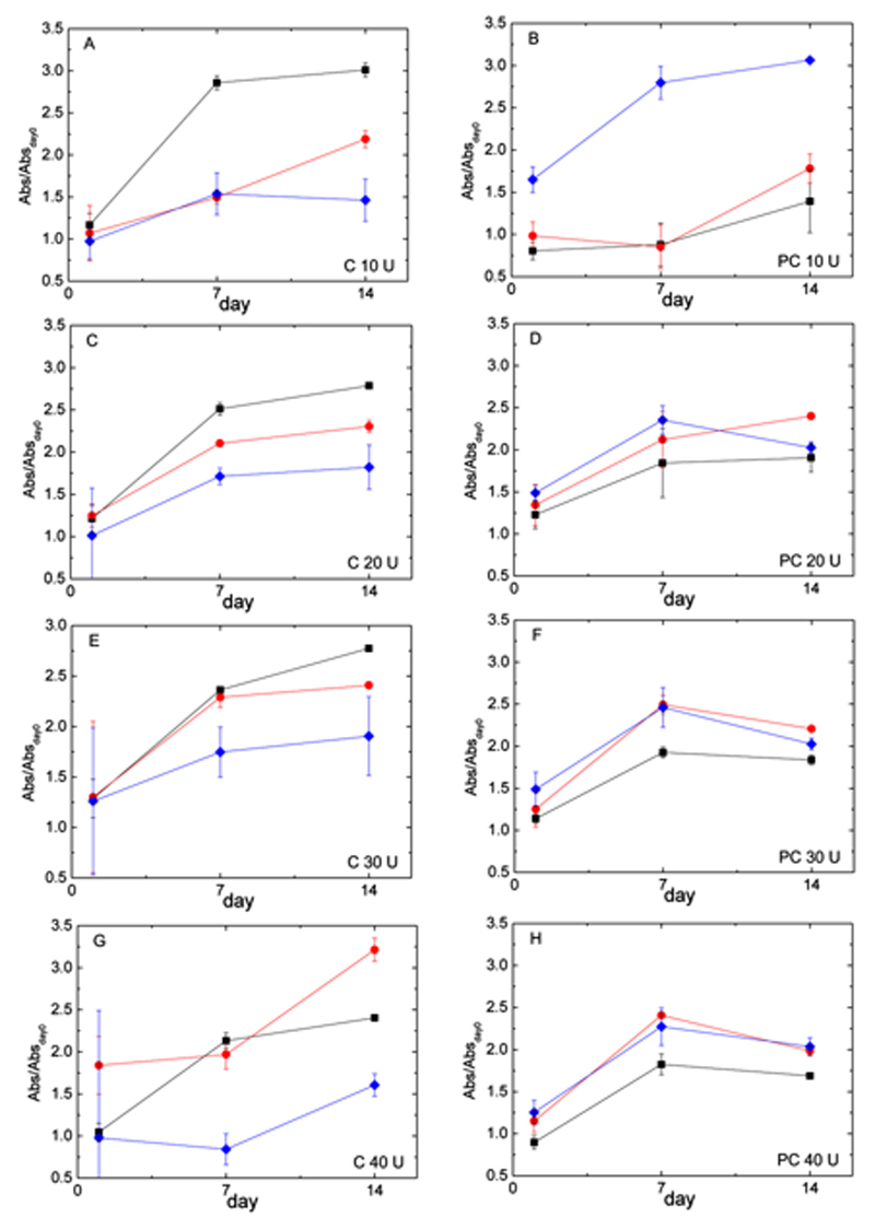
Figure 7.
Cell proliferation of MC3T3 cells on hydrogels. Cells were cultured on Gel 10% (▪), Cs 0.10%/Gel 10% (●) and Cs 0.50%/Gel 10% (♦) (wt%). Data were collected at 1, 7 and 14 days using an MTS assay (normalized to day 0) for chemical (A, C, E, G) and physical-co-chemical hydrogels (B, D, F, H) at A,B) 10, C,D) 20, E,F) 30, and G,H) 40 U/ggelatin of transglutaminase. Values are mean ± SD.

