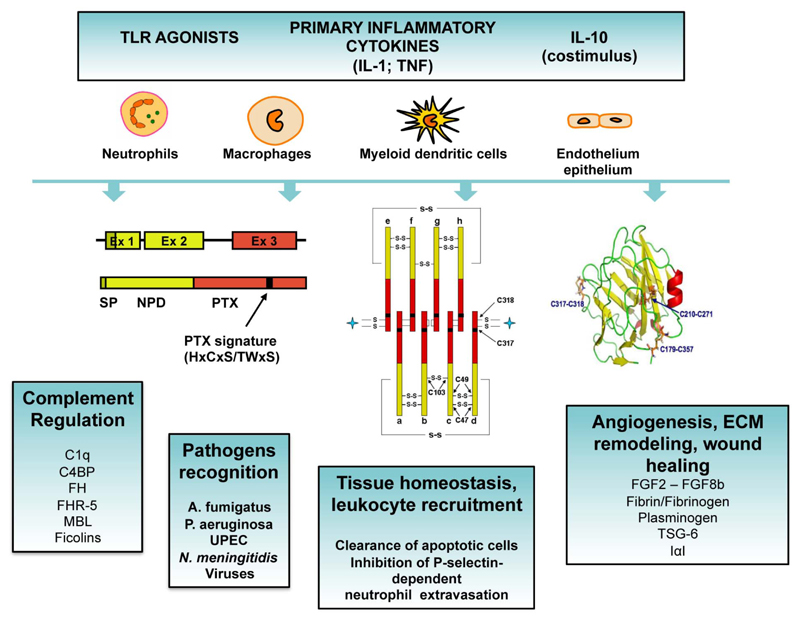
Figure 2. Expression, protein structure and roles of PTX3.
Inflammatory cytokines, TLR agonists and microbial moieties induce the expression of PTX3 in various cell types (i.e. dendritic cells, monocytes, macrophages, epithelial cells, endothelial cells, fibroblasts and adipocytes). Neutrophils store PTX3 in specific granules in a ready-to-use form. The gene coding for PTX3 is organized in three exons. The first two exons code for the signal peptide (SP) and the N-terminal domain of the protein (NTD), respectively, and the third exon codes for the pentraxin domain (PTX) containing the PTX signature. PTX3 has a quaternary structure with eight subunits, associated together to form an octamer by a network of disulfide bonds. The three-dimensional model of the pentraxin domain has been generated based on the crystallographic structures of CRP and SAP, showing that the pentraxin domain of PTX3 adopts a β-jelly roll topology. PTX3 plays a role in complement activation and regulation, pathogen recognition, leukocyte recruitment, recognition of apoptotic cells, angiogenesis, extracellular matrix (ECM) remodelling and wound healing.

