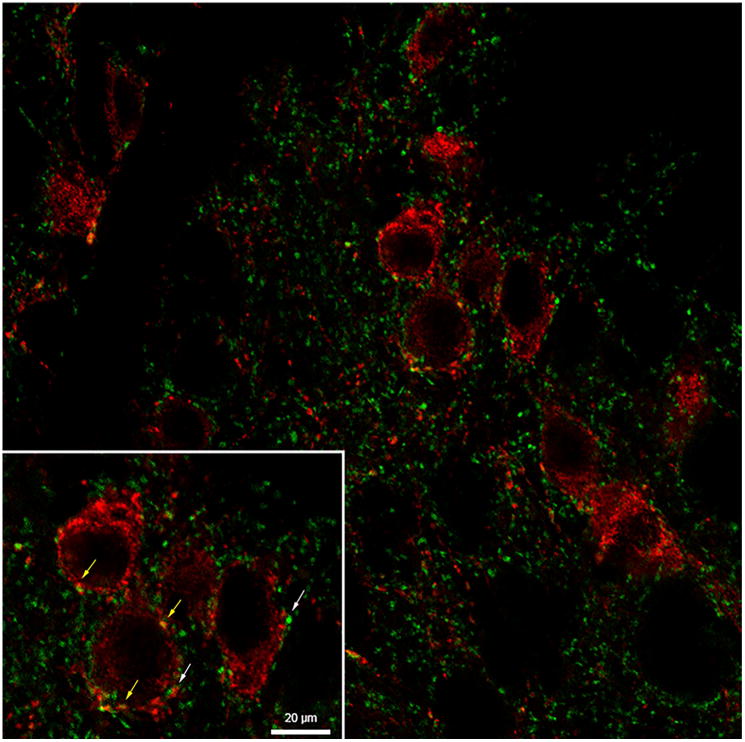
Figure 6.
Representative image of kisspeptin-immunopositive neurones (red) with synaptophysin-positive close-contacts (green) within the arcuate nucleus from an ovariectomized prepubertal lamb. Synaptophysin to kisspeptin contacts are denoted by white arrows and kisspeptin to kisspeptin contacts denoted by yellow arrows. Scale bar = 20 μm (inset).

