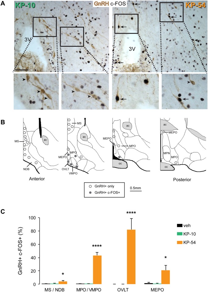
Fig 2. Peripheral administration of KP-54 but not KP-10 induces c-FOS in GnRH neurons.
(A) Representative photomicrographs showing coronal sections of dual-labelled immunohistochemistry showing c-FOS staining (black nuclei) and GnRH neurons (brown cells) at the level of the OVLT and MPO regions in the mouse hypothalamus 2 hrs after peripheral injection of KP-54 (n = 6) or KP-10 (n = 6). GnRH neurons with c-FOS staining are indicated with black arrows. Grey arrowheads indicate c-FOS negative GnRH neurons. KP-54 (but not KP-10) stimulates c-FOS in the hypothalamus (and this co-localises with GnRH neurons). Scale bar = 20 μm. (B) Schematic diagrams showing distribution of c-FOS positive (closed circles) and c-FOS negative (open circles) GnRH neurons throughout the anterior hypothalamus following peripheral administration of KP-54 (n = 6). Diagrams are re-drawn from those available from the Allen Institute for Brain Science (www.alleninstitute.org/). ac anterior commissure; MEPO, median preoptic nucleus; MPN, medial preoptic nucleus; MPO, medial preoptic nucleus; MS, medial septum; NDB, diagonal band nucleus; oc, optic chiasm; OVLT, organum vasculosum of lamina terminalis; VMPO, ventromedial preoptic nucleus. (C) Histogram showing the number of GnRH neurons expressing c-FOS 2 hrs after PBS (veh, n = 6), KP-54 (n = 6) or KP-10 (n = 6) in different hypothalamic regions. MS/NDB, medial septum and diagonal band nucleus; MPO/VMPO, medial preoptic area and ventromedial preoptic nucleus; OVLT, organum vasculosum of lamina terminalis; MEPO, median preoptic nucleus. Significant differences between KP-54 and vehicle or KP-10 are indicated (one-way ANOVA, followed by Tukey’s multiple comparison test).

