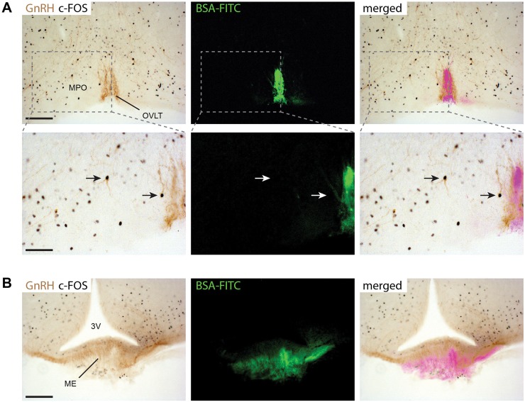
Fig 3. Peripheral administration of KP-54 activates GnRH neurons inside the blood-brain-barrier in male mice.
KP-54 (1nmol) was injected into male mice (n = 3) and activation of c-FOS in GnRH neurons determined after 2 h (A) Dual-label immunohistochemistry showing GnRH neurons (brown cytoplasmic staining) and c-FOS staining (black nuclei) in the OVLT/MPO region. Low-power view (upper panels) and higher magnified views (lower panels) of the dotted line boxes show c-FOS + GnRH neurons (arrows) in the OVLT and MPO hypothalamic regions. The c-FOS + GnRH neuron in the MPO (arrow in the centre of the picture) is clearly located inside the blood brain barrier defined by BSA-FITC staining. Scale bars: 100 μm (upper panels) and 50 μm (lower panels). (B) GnRH axon projections (brown) overlap with the BSA-FITC staining in the median eminence (EM) as this region is devoid of a blood-brain-barrier. Scale bar = 100 μm. 3V: third ventricle, OVLT: organum vasculosum of lamina terminalis, MPO: medial preoptic area.

