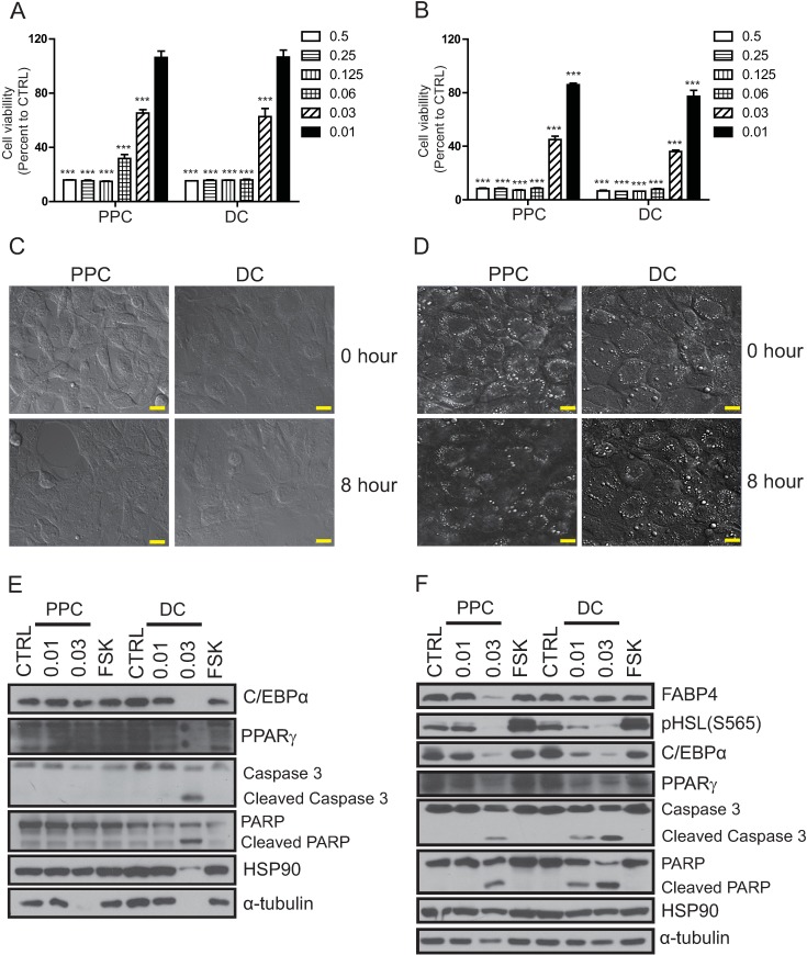
Fig 2. Effect of phosphatidylcholine (PPC) or deoxycholate (DC) on the viability of preadipocytes and adipocytes.
A. 3T3L1 preadipocyte and B. adipocyte viability was measured eight hours after treatment with various doses of PPC or DC using the MTT assay. Live confocal images of C. 3T3L1 preadipocytes and D. adipocytes captured before (0 h) and eight hours of treatment with 0.03% of PPC or DC. The white dots indicate the lipid vacuoles of mature adipocytes, and the eight-hour treatment with PPC decreased the number of lipid vacuole-positive cells to a greater extent than DC treatment. Protein samples from E. 3T3L1 preadipocyte and F. adipocytes treated with 0.01% or 0.03% of PPC or DC respectively for eight hours, were prepared and the expression of preadipocyte (C/EBPα, PPARγ, and HSL), and mature adipocyte (C/EBPα, PPARγ, HSL, and FABP4) markers, apoptotic markers (cleaved-caspase3 and cleaved-parp), as well as the amount of loading controls (HSP90 and α-tubulin) were analyzed using Western blotting. The scale bar indicates 20 μm.

