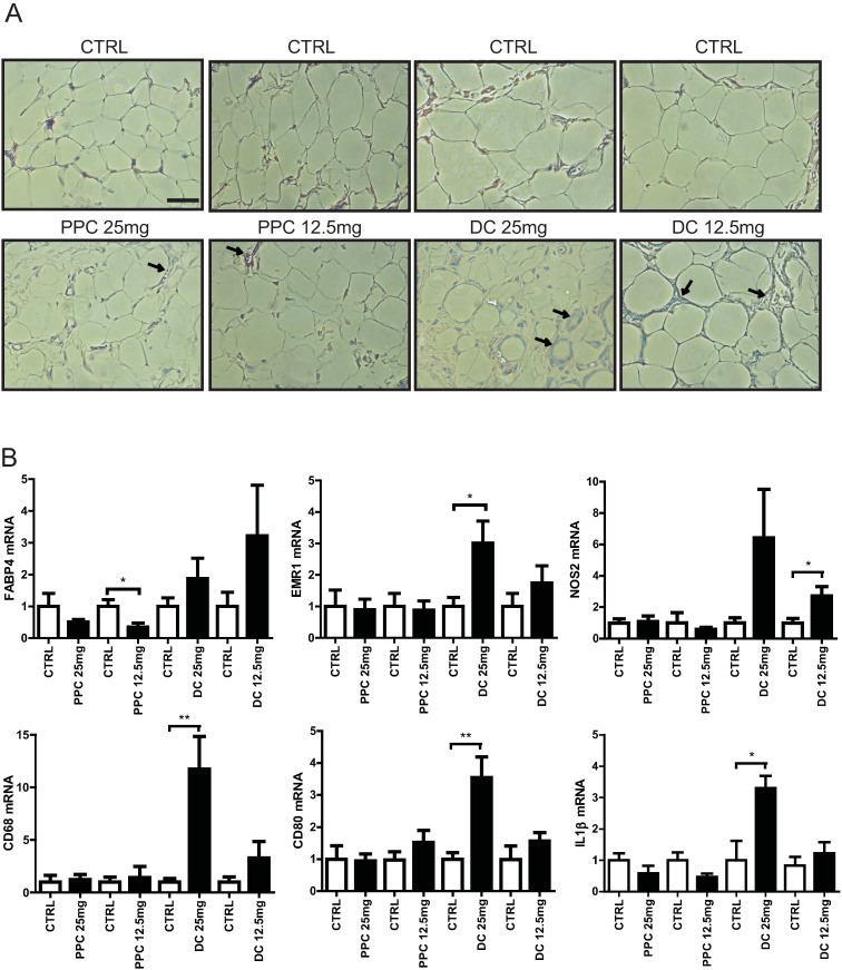
Fig 5. Effects of phosphatidylcholine (PPC) and deoxycholate (DC) on inguinal adipose tissue in the rat.
A. Hematoxylin and eosin staining of inguinal adipose tissue sections from rats 30 days after control, PPC, or DC injection. The arrow indicates macrophage infiltration and the scale bar indicates 50 μm. B. Gene expression analysis of inguinal adipose tissue injected with control or 25 mg or 12.5 mg of PPC or DC, respectively. Using real-time qRT-PCR, the relative expression levels of FABP4, a marker for mature adipocytes and EMR1, NOS2, CD68, CD80, and IL1β markers for macrophages, in inguinal adipose tissue injected with PPC or DC were compared with the expression in inguinal adipose tissue injected with ethanol or PBS control solutions, respectively.

