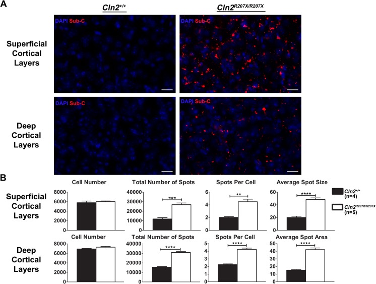
Fig 8. Lysosomal accumulation of mitochondrial ATP synthase subunit c in Cln2R207X/R207X mice.
Subunit c accumulation was detected by immunofluorescent staining. (A) Images of superficial and deep cortical layers demonstrate diffuse and pronounced accumulation of mitochondrial ATP synthase subunit c in 3-month-old Cln2R207X/R207X mice. (B) Images from 3-month-old Cln2+/+ (n = 4) and Cln2R207X/R207X (n = 5) mice were blindly collected and analyzed for differences in cell number, total number of immunoreactive puncta, number of puncta per cell, and average punctum area. Cln2R207X/R207X mice have significantly increased total number of puncta, puncta per cell, and punctum size. Columns and bars represent mean ± SEM. Statistical significance was determined using an unpaired t-test (**p < 0.01, ***p < 0.001, and **** p < 0.0001).

