Figure 1.
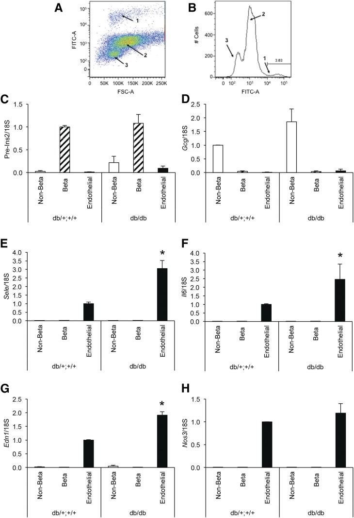
(A, B) Separation of islet cells from 8-week-old db/db and db/+;+/+ mice by fluorescence-activated cell sorting, yielding fluorescently labeled endothelial cells (population 1), β cells (population 2), and non–β-cells (population 3), the latter two differentiated by autofluorescence. (C–H) mRNA levels determined by quantitative polymerase chain reaction in the islet non–β (open bars), β (hatched bars), or endothelial (solid bars) cell populations from db/db or db/+;+/+ littermate controls. Expression of Ins2 pre-mRNA (C) and Gcg mRNA (D) demonstrates good separation among the cellular populations (high in β cells and non–β cells, respectively, and low in the other populations). Endothelial markers Sele (E), Il6 (F), Edn1 (G), and Nos3 mRNA (H) show selective expression in islet endothelial cells. Data are presented as mean ± standard error of the mean; n = 5 per genotype group; *P < 0.05 vs db/+;+/+ endothelial cells.

