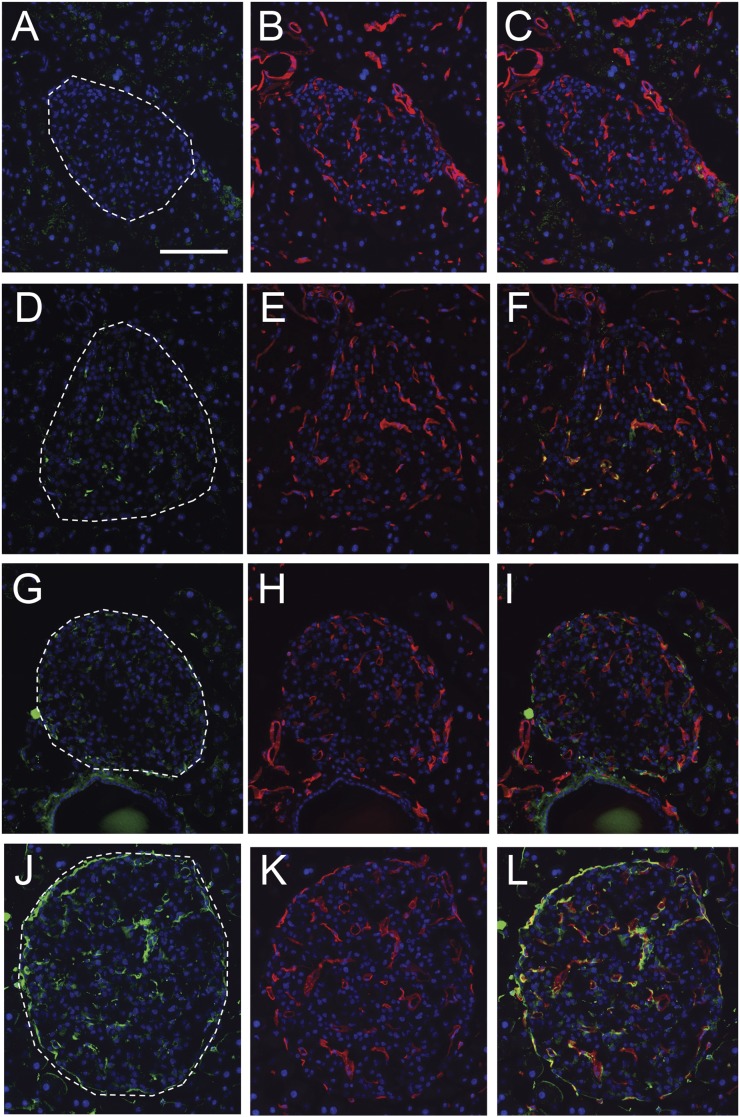
Figure 4.
Representative micrographs of pancreas showing immunoreactivity for AGE (green, A, D, G, J) and CD31 to visualize islet endothelial cells (red; B, E, H, K) with nuclear counterstaining (blue; all panels). Merged images showing colocalization between these two markers (orange or yellow staining) or close apposition thereof (C, F, I, and L). AGE immunoreactivity was absent from islets from 8-week-old db/+;+/+ control mice (A–C) and from those of 16-week db/+;+/+ control mice (G–I). In contrast, AGE immunoreactivity was observed exclusively in CD31-positive cells in 8-week-old db/db diabetic mice (G–I). More extensive AGE immunoreactivity was observed in islets from 16-week-old db/db mice (J–L), all of which was localized within or closely associated with CD31-positive cells. Scale bar = 100 µm.

